Terahertz Imaging Advances Diagnostics
Terahertz Imaging is emerging as a beacon of innovation in non-invasive medical diagnostics, marrying the subtle science of sub‑millimeter wavelengths with the need for rapid, radiation‑free assessment of human tissues. This technology sits where optical imaging, ultrasound, and MRI converge, yet it offers a unique spectral window that can detect water content, mineral density, and biochemical signatures essential for early disease detection. In what follows, we explore the science behind Terahertz Imaging, its clinical promise, its distinct advantages, and the hurdles it faces before becoming mainstream.
Terahertz Imaging: Foundation of Non-Invasive Diagnostics
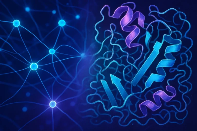
The terahertz (THz) portion of the electromagnetic spectrum, spanning 0.1–10 THz, is often referred to as the “missing link” between microwaves and infrared light. Thanks to their photon energy, THz waves are non-ionizing, making them inherently safer for medical use. They interact with polar molecules such as water and hydrogen bonds, revealing subtle differences in tissue composition. For instance, the dielectric contrast between healthy and cancerous skin layers can be quantified, enabling high‑contrast imaging without the need for invasive contrast agents.
For a deeper dive into the physics, the Wikipedia terahertz radiation page provides foundational insight into their generation and detection mechanisms. Medical researchers use gyrotrons, quantum cascade lasers, and optical rectification to produce coherent THz beams, while photoconductive antennas or bolometers capture the returning signal.
Clinical Applications: From Oral Health to Oncology
Research projects worldwide have demonstrated the utility of Terahertz Imaging in multiple settings:
- Detecting early enamel demineralization in dental clinics, allowing preventive treatments before cavities form.
- Mapping bone density changes in osteoporosis patients, offering a radiation‑free alternative to DXA scans.
- Identifying breast tissue architecture abnormalities in high‑risk individuals, potentially reducing unnecessary biopsies.
- Providing rapid intraoperative feedback during tumor resections, helping surgeons ensure clear margins while sparing healthy tissue.
Clinical writers such as the Mayo Clinic have reported on pilot studies where THz scans successfully distinguished malignant lesions from benign ones by their water absorption spectra. These trials underscore the potential for Terahertz Imaging to supplement, or in some cases replace, more invasive diagnostic procedures.
Advantages Over Conventional Modalities
While ultrasound, CT, and MRI each have advantages, Terahertz Imaging offers several key benefits that align well with contemporary patient care priorities:
- Non-ionizing radiation: Unlike X-rays and CT, THz waves pose no risk of DNA damage, making them suitable for frequent use and for vulnerable populations such as children and pregnant patients.
- Real‑time imaging: Modern detectors can capture data at rates exceeding 10 frames per second, allowing clinicians to monitor dynamic processes such as blood flow or tissue hydration in vivo.
- Label-free contrast: By exploiting intrinsic molecular vibrations, THz imaging eliminates the need for external dyes or agents, simplifying workflow and reducing potential allergic reactions.
- High depth resolution: THz waves can penetrate several millimetre depths in soft tissues, placing them in a middle ground between optical surface imaging and deeper ultrasound imaging.
- Cost‑effective hardware: Recent developments in photonic sources have dramatically reduced the price point of THz systems compared to full‑scale MRI scanners.
Challenges and Future Directions
Despite the promise, several technical and regulatory challenges remain before Terahertz Imaging can be broadly applied:
- Limited penetration depth: In highly scattering tissues, THz signals can degrade, limiting clear imaging to superficial layers unless enhanced detectors or adaptive optics are employed.
- Atmospheric humidity: Water vapour strongly absorbs terahertz frequencies, necessitating dry or controlled environments during data acquisition.
- Standardization and calibration: Unlike Raman or MRI, industry‑wide calibration standards for THz scanners are still under development, making cross‑study comparison difficult.
- Regulatory approval: Organizations such as the U.S. Food & Drug Administration are currently evaluating devices that integrate THz imaging, and clinical trials must demonstrate safety and efficacy before commercial deployment.
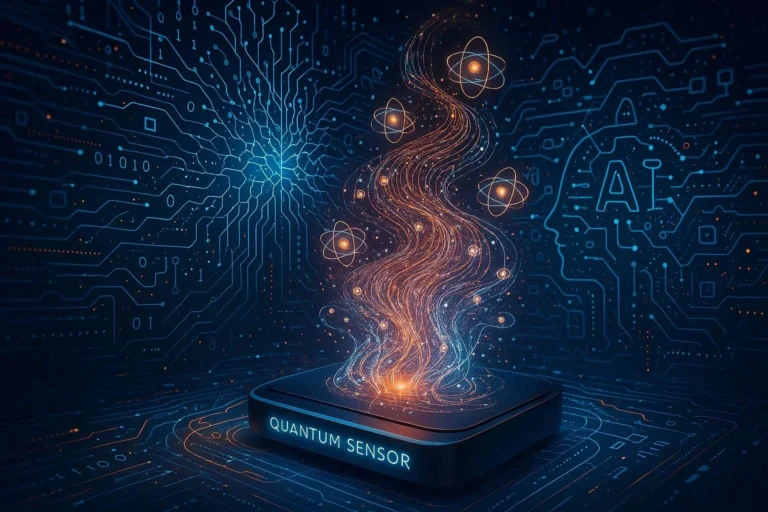
Looking ahead, collaborations between academia and industry are spurring the emergence of ultra‑high‑speed, miniature THz emitters powered by deep learning algorithms that reconstruct images from fewer frames. The NIH recently funded a consortium aimed at linking terahertz spectra to specific tumor signatures, which could lead to rapid point‑of‑care diagnostics in future operating rooms.
Emerging Research Trends
Recent pre‑clinical studies indicate that composite imaging—combining terahertz with optical or ultrasound data—can enhance diagnostic accuracy. Additionally, machine‑learning models trained on terahertz datasets are showing remarkable ability to classify early-stage pancreatic lesions from benign cysts, a notoriously challenging area in oncology.
In Conclusion
Terahertz Imaging stands poised to revolutionize non-invasive medical diagnostics by marrying safety, speed, and molecular specificity. While obstacles remain, ongoing research, regulatory attention, and strategic investments suggest that within the next decade we may witness widespread deployment of THz systems in dermatology, dentistry, orthopedics, and oncology. If you’re a clinician seeking cutting‑edge diagnostic tools, or a researcher passionate about biophotonics, now is the perfect time to engage with terahertz technologies. Discover how terahertz imaging can elevate patient care by contacting our team for a personalized demo and roadmap today.
Frequently Asked Questions
Q1. What is Terahertz Imaging?
Terahertz Imaging uses electromagnetic waves between microwave and infrared ranges to visualize tissue composition without ionizing radiation. It captures subtle differences in water content and molecular bonds, enabling high‑contrast imaging of healthy versus diseased tissue.
Q2. How does it differ from other imaging modalities?
Unlike X‑ray, CT, or MRI, terahertz signals are non‑ionizing and offer intrinsic contrast based on molecular vibrations. It also penetrates several millimetres into soft tissue, providing a depth resolution that falls between optical imaging and ultrasound.
Q3. What clinical advantages does Terahertz Imaging provide?
It delivers real‑time, label‑free scans that are safe for frequent use, especially in children and pregnant patients. Clinical trials show early detection of enamel demineralization, bone density changes, and breast tissue abnormalities with minimal patient discomfort.
Q4. What are the main technical challenges?
Key obstacles include limited penetration depth in highly scattering tissues, strong absorption by atmospheric water vapor, and the need for industry‑wide standardization and calibration protocols.
Q5. Is Terahertz Imaging safe for pregnant patients and children?
Yes—since terahertz waves are non‑ionizing, they pose no known risk of DNA damage. Clinical guidelines emphasize using the lowest effective dose and ensuring patient comfort to maintain safety during scans.
Related Articles

100+ Science Experiments for Kids
Activities to Learn Physics, Chemistry and Biology at Home
Buy now on Amazon
Advanced AI for Kids
Learn Artificial Intelligence, Machine Learning, Robotics, and Future Technology in a Simple Way...Explore Science with Fun Activities.
Buy Now on Amazon
Easy Math for Kids
Fun and Simple Ways to Learn Numbers, Addition, Subtraction, Multiplication and Division for Ages 6-10 years.
Buy Now on Amazon